Anatomy BioArt Winners

Winners were chosen by a panel of AAA members from all anatomy entries to the FASEB BioArt Scientific Image & Video Competition and announced during Virtual Annual Meeting Week, April 6-10, 2020.

Image by Caitlin Yoakum

Caitlin Yoakum, University of Arkansas

This is a microCT image of a juvenile squirrel monkey (Saimiri sciureus). This monkey has all of its deciduous (baby) teeth in place, but its adult teeth have formed just below them, waiting to erupt as the baby teeth fall out.

Image by Akinobu Watanabe

Akinobu Watanabe, New York Institute of Technology College of Osteopathic Medicine

Forging a New Path: The image was taken after grafting Hensen’s node (organizer) from an early-stage chicken embryo labeled with green fluorescence next to the host embryo (bottom left), inducing a new embryo to develop away from the host.

Image by Susan Motch Perine et al.

Susan M. Motch Perrine, Joan T. Richtsmeier, & Kazuhiko Kawasaki, The Pennsylvania State University; Danny Chen & Hao Zheng, University of Notre Dame

Depicted is automatically segmented cartilage and skin from a microCT scan of a mouse at embryonic day 14.5. This image is used to examine how early cartilaginous structures serve as a structural and functional scaffold for development of the head.

Image by Adam Hartstone-Rose & Edwin Dickinson

Adam Hartstone-Rose & Edwin Dickinson, North Carolina State University

The first fascicle-by-fascicle digital dissection of lemur masticatory muscles depicting those of a typical lemur (a mongoose lemur, top) and the enigmatic aye-aye (bottom).

Masseter = green; temporalis = red; pterygoid = blue/purple.

Image by Brent Adrian et al.

Brent Adrian, Heather F. Smith, Avery Williams, Aryeh Grossman, & Andrew Lee, Midwestern University; Christopher Noto, University of Wisconsin-Parkside

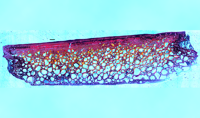
Paleohistological thin section from a 96 million year old fossil pond turtle shell. Polarized light reveals changes in orientation patterns of collagen fibers in the bone interior. Also, vascular channels and growth marks are visible.

Named a FASEB BioArt Winner, December 2019.